A skinfold measurement estimates your body fat percentage from the thickness of your subcutaneous fat at three to seven sites. In practised hands it lands within three to five percent. Better than a home scale, worse than a DEXA scan, for about twenty euros of equipment.
The largest source of error is not the caliper. It is whoever holds it.
I measured myself weekly for half a year before my figures settled. That learning curve is part of the method, and nobody mentions it.
What does a skinfold measurement actually measure?
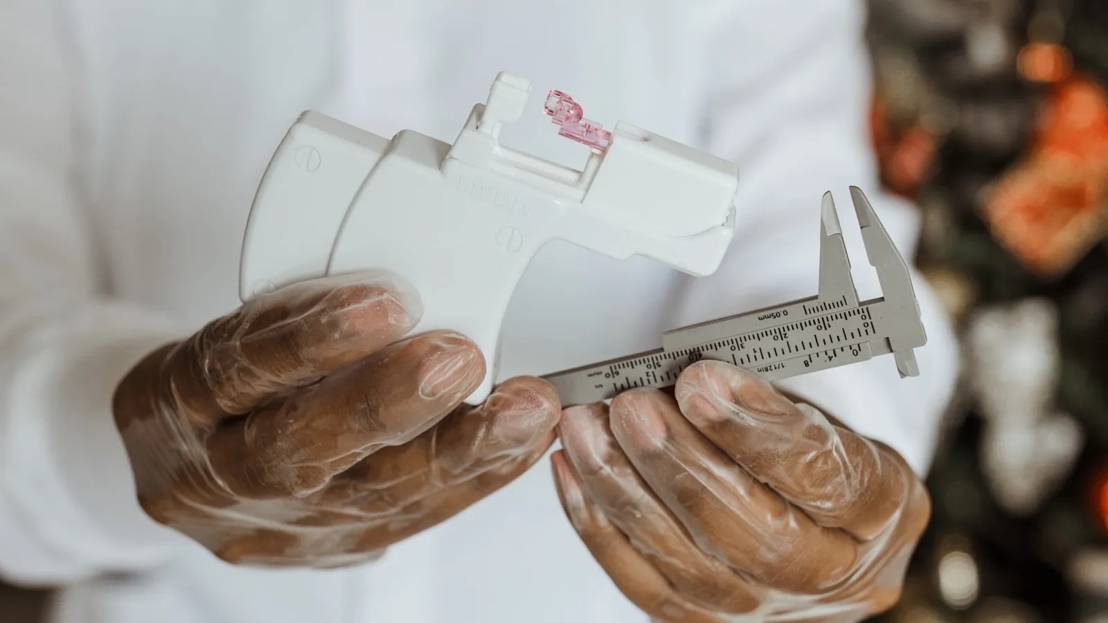
Fat tissue directly under your skin, and nothing else. You pinch a fold of skin plus subcutaneous fat between thumb and forefinger, place the caliper a centimetre below, and read the thickness in millimetres after two seconds. Those millimetres then go through an equation.
So what the caliper never sees is the fat around your organs.
That is a real limitation. Two men with identical skinfolds can differ substantially in abdominal fat, and that fat is the one most strongly linked to metabolic problems. We cover it in visceral fat.
A skinfold measurement is therefore a fine trend tool and a mediocre health measure.
Which sites do you measure?
That depends on the protocol. The best known come from Jackson and Pollock: a three, four or seven-site measurement, with different sites for men and women. More sites cost more time and give a slightly steadier figure, because one badly taken fold carries less weight.
| Protocol | Sites in men | Sites in women | In practice |
|---|---|---|---|
| 3-site | chest, abdomen, thigh | triceps, suprailiac, thigh | quick, fine at home |
| 4-site | triceps, biceps, subscapular, suprailiac | same four sites | most used in Dutch gyms |
| 7-site | the three plus triceps, subscapular, suprailiac, midaxillary | the three plus chest, subscapular, midaxillary | steadiest, needs practice |
Pick one and stay with it. Switch protocol and you switch equation, which makes your old figure incomparable.
Always measure on the right side, always standing, always before training. Take each site three times; if the third differs by more than two millimetres, measure again.
Why is your calculated percentage not exact?
Because two translation steps sit inside it. The Jackson and Pollock equations do not predict your body fat percentage but your body density (PMID 718832, and the 1980 version for women, PMID 7402053). Only a second equation, usually Siri or Brozek, converts that density into a percentage of fat.
Each step carries its own error. They stack.
Those equations were also derived in the 1970s from samples that may look nothing like you. If you are heavily muscled, past sixty, or of non-European descent, you sit further from the group the formula was calibrated on.
The IOC working group around Ackland put it plainly in 2012: no practical method measures fat directly, and every field method inherits the assumptions of the reference method it was calibrated against (PMID 22303996). Skinfolds are the textbook case.
That matters less than it sounds, as long as you use the measurement as a trend. Take a four-site measurement dropping from 42 to 34 millimetres across eight weeks: you are losing fat, whichever equation you run it through.
The formula may sit two percent off in absolute terms. As long as it is off by the same amount every time, the direction still holds.
That is how I use it myself. Not to answer how fat I am, but which way it is going.
How do you avoid the common mistakes?
By keeping the measurement dull and identical. The four errors that wreck your figure hardest are: measuring at the wrong spot, pinching too loose or too tight, reading too quickly, and switching testers. That last one costs more accuracy than any caliper will ever give back.
Two measurements by the same person beat one measurement by an expert.
Practically: mark the sites with a pen, read two seconds after releasing the caliper, and keep holding the fold while you read. Ideally let someone else take your back and triceps sites, because you cannot pinch those reliably yourself.
Do not measure straight after cardio or a sauna either. Blood flow and fluid distribution in the skin change fold thickness temporarily.
Skinfolds or another device?
Choose on what the measurement has to carry. For a trend every six weeks, skinfolds beat a scale and cost far less than a scan. If you want an absolute figure to base a long cut on, that is the point to consider a DEXA scan.
The comparison across all five methods, error margins side by side, is in body fat percentage: what is healthy and how to measure it. Why bioimpedance swings so much in trained people is in the body composition scale.
Skinfolds have one advantage that gets forgotten: they are insensitive to your fluid balance. Where a scale can read two percent higher after a low-carb week, your folds stay where they were.
What the caliper does not measure is whether your fat tissue is getting in your metabolic way. According to figures RIVM tracks through the Leefstijlmonitor, about half of Dutch adults carry excess weight, and the metabolic picture inside that group varies enormously.
My advice: use the caliper for the direction of your fat loss and let your lipids answer the metabolic question. Every blood test result includes a professional assessment from a BIG-registered doctor. For treatment decisions, discuss your results with your GP.
References
- Jackson AS, Pollock ML. Generalized equations for predicting body density of men. British Journal of Nutrition, 1978;40(3):497-504. PMID 718832.
- Jackson AS, Pollock ML, Ward A. Generalized equations for predicting body density of women. Medicine and Science in Sports and Exercise, 1980;12(3):175-81. PMID 7402053.
- Ackland TR, Lohman TG, Sundgot-Borgen J, et al. Current status of body composition assessment in sport. Sports Medicine, 2012;42(3):227-49. PMID 22303996.
- Esco MR, Snarr RL, Leatherwood MD, et al. Comparison of total and segmental body composition using DXA and multifrequency bioimpedance in collegiate female athletes. Journal of Strength and Conditioning Research, 2015;29(4):918-25. PMID 25353076.
- RIVM. Leefstijlmonitor, figures on overweight in adults.
الكاتب